OUR PRACTICE
Welcome to Westside Endodontics in Calgary, Alberta. Our focus is on delivering high-quality root canal treatment, root canal retreatment, and root surgery in a calm, caring manner. We take great pride in the partnerships we’ve developed with leading dentists to deliver optimal dental care. As proud Calgary Endodontists, we’re immensely happy to have helped numerous Calgarians improve their dental health and preserve their teeth for a lifetime.
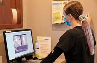
ADVANCED TECHNOLOGY
DIGITAL RADIOGRAPHS
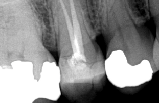
Dental radiographs taken at your dental office are used to look for dental decay and to better view restoration margins. The radiographs we take at Westside Endodontics are done at different angles (than the ones taken at your dental office) to allow better visualization of overlapping roots. This is why we may require additional radiographs, even though it seems your dentist already took images of the same tooth.
We use digital imaging at Westside Endodontics, which has much less exposure time than traditional radiographs, resulting in 80% less radiation exposure to our patients. Digital images are also easier to store and transfer (when needed).
MICROSCOPES
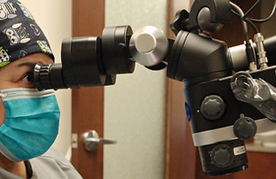
During root canal therapy, magnification and illumination provided by the operating microscope aids in numerous ways. The additional magnification and light help with:
-
Identifying complex canal anatomy.
-
Seeing cracks and fractures not visible to the naked eye.
-
Localization of obstructed and calcified canals.
-
The removal of canal obstructions such as denticles and calcifications.
-
Treatment of dental anomalies, such as dens invaginatus, or fused teeth.
-
In endodontic retreatments, the microscope is helpful in identifying and removing leftover filling materials.
-
Surgical endodontics has been completely transformed by microscopic procedures. Classic root surgeries are now microsurgeries, using smaller instruments to help reduce the amount of tooth structure removed.
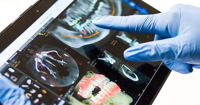
CBCT
Cone-beam computed tomography is an innovative imaging technique that provides endodontists with three-dimensional views inside of the patient’s tooth. A CBCT greatly enhances the endodontist’s ability to diagnose, evaluate and treat complex canal anatomy, canal pathology and get a clearer idea of the internal anatomy of a patient’s tooth before treatment is initiated.
During a CBCT scan, the machine rotates around the patient, capturing images using a cone-shaped X-ray beam. These images are then used to construct a 3-D representation of the patient’s teeth and oral and maxillofacial region (teeth, jaw bone, and the temporomandibular joint). For endodontic treatment, we use a limited volume CBCT that shows exceptional detail but is focused only on a few teeth.
CBCT images are taken when there are complex endodontic conditions, such as:
-
Root Canal system anomalies & to determine root curvature
-
Diagnosis of dental root infections in patients who present with nonspecific clinical signs and symptoms or no evidence of disease visible in conventional imaging.
-
To assess prior treatment complications, such as overextended root canal obturation material, separated endodontic instruments, calcified canal identification, and localization of perforations.
-
Diagnosis and management of dental trauma, especially root fractures, luxation and/or displacement of teeth, and alveolar fractures.
-
Presurgical case planning to determine the exact location of root apex/apices and to evaluate the proximity of adjacent anatomical structures, such as nerves.
CBCT images are also taken prior to placement of dental implants to:
-
Perform presurgical case planning for implant placement
-
To determine the location of important anatomical structures including the roots of teeth that will be adjacent to the implant and the location of nerves.